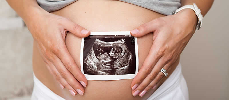
Throughout the history, pregnant women routinely undergo ultrasound examination, a test that uses high-frequency sound waves to image the developing baby and the mother’s placenta and uterus. This is done during the second trimester of the pregnancy, usually between 18 and 20 weeks, as a check to determine that all things are going as they should.
This examination is usually a happy moment and the parents often come away with their “first selfie” of their new baby. The ultrasound, however, has a very important diagnostic function which can provide critical information to the new parents regarding the health of their baby.
An ultrasound or a sonogram can help monitor normal fetal development and screen for any potential problems. Along with a standard ultrasound, there are few more advanced ultrasounds, including a 3D ultrasound, a 4D ultrasound, and a fetal echocardiography, which is an ultrasound that gives a detailed look at the fetus’ heart.
An ultrasound can be used for a variety of reasons during pregnancy. Your doctor may also order more ultrasound examinations if they detected a problem in a previous examination or blood test. Ultrasounds may also be done for nonmedical reasons, such as to produce images for the parents or to determine the sex of the baby.

On its most basic level, ultrasound imaging is used to assess a developing pregnancy for the following:
- Confirm the pregnancy
- Gestational age
A normal pregnancy is popularly thought of as 40 weeks gestation. However, in medical terms, a term pregnancy is anywhere from 37 to 41 weeks. It is important to verify the gestational age of the developing fetus for several reasons. For example, the growth of the baby will be measured against well-established growth charts to ensure normal development. Gestational age will be verified against the dates provided by the mother regarding her last menstrual period to confirm the due date and ensure that the baby is not delivered either too early or too late.
- Check for multiple pregnancies (twins, triplets, etc)
A pregnancy with multiple babies carry special risks and must be monitored on a regular basis. Complications such as a “twin to twin transfusion” and cervical incompetence require prompt attention if complications are to be avoided.
- Problems with the placenta
During pregnancy the position of the placenta within the uterus can be vitally important to the health of both the baby and, in some circumstances, the mother. An ultrasound can determine complications such as: placenta previa, vasa previa, placenta accrete, placenta increta, and placenta percreta.
- Monitor fetal position
During delivery it can be important to know the baby’s position because it can affect the method of delivery.
- Check for congenital anomalies
Many parents will want to know if their baby suffers from any congenital or genetic problems, so they can terminate the pregnancy or prepare for the difficulties associated with the particular problem.
- Monitor fetal growth
If the growth of the baby falls off expected norms this can be indicative of problems with the placenta or problems with the health of the baby. Either way, early intervention may be required in order to deal with the problem.
- Monitor the level of amniotic fluid
Amniotic fluid is produced by the fetus. Too much or too little amniotic fluid may be indicative of problems with pregnancy which may require intervention.
Different types of ultrasound imaging are used for different purposes. The different kinds of ultrasound available include:
- Transabdominal ultrasound
When you hear about ultrasound during pregnancy, it’s most likely this kind. You lay on your back on an exam table, and your obstetrician covers your belly with a thin layer of gel. The gel helps the sound waves move more easily so you get a better picture. Ultrasound is painless but having a full bladder may be uncomfortable. The ultrasound takes about 20 minutes.
- Transvaginal ultrasound
This type of ultrasound is done through the vagina. You lay on your back on an exam table with your feet in stirrups. Your obstetrician moves a thin transducer into your vagina. You may feel some pressure from the transducer, but it shouldn’t cause pain. The procedure takes about 20 minutes.
- 3D ultrasound
A 3D ultrasound takes thousands of pictures at once. It makes a 3D image that’s almost as clear as a photograph. Some providers use this kind of ultrasound to make sure your baby’s organs are growing and developing normally. You also may get a 3D ultrasound to check for problems in the uterus.
- 4D ultrasound
This is like a 3D ultrasound, but it also shows your baby’s movements in a video.
During an ultrasound earlier in the pregnancy full bladder for the doctor to get a clear image of the fetus and your reproductive organs. You should drink two to three glasses of water one hour prior to your scheduled ultrasound. You shouldn’t urinate before the examination, so you arrive at your appointment with a full bladder.
Ultrasound is safe for you and your baby when done by healthcare professionals – Obstetricians or a diagnostic medical sonographer. Because ultrasound uses sound waves instead of radiation, it’s safer than X-rays. Providers have used ultrasound for more than 30 years, and they have not found any dangerous risks.
If your pregnancy is healthy, ultrasound is good at ruling out problems, but not as good at finding them. It may miss some birth defects. Sometimes, a routine ultrasound may suggest that there is a birth defect when there really isn’t one. While follow-up tests often show that the baby is healthy, false alarms can cause worry for parents.
For most women, ultrasound shows that the baby is growing normally. If your ultrasound is normal, just be sure to keep going to your prenatal checkups.
Sometimes, ultrasound may show that you and your baby need special care. No matter what an ultrasound shows, talk to your doctor about the best care for you and your baby.
For more information, watch the video below.
References:


