


By continuing to browse our site, you agree to the storing of cookies on your device to help us understand your preferences and improve our services, ensuring that we can provide you with the best possible experience. To learn more, check out our Cookie Policy.
- ABOUT US
- Medical Specialty Care
- OUR SERVICES
- PARTNERS
- IDSMED LEARNING ACADEMY
- InnoQ
- News
- Talent & Jobs
- LOG IN / SIGN UP
+62 21 2567 8989
- English change
- Home
- About Us
-
Medical Specialty Care
- Aesthetic
- Anesthesiology & Analgesia
- Bio-Medical Services
- Cardio Vascular
- Dental
- Diagnostic Imaging
- Ear, Nose & Throat
- Emergency Care
- Gastroenterology
- General Surgery
- Geriatric Medicine
- Healthcare Education
- Infection Control
- Intensive Care
- Laboratory
- Medical Consumables
- Medical IT
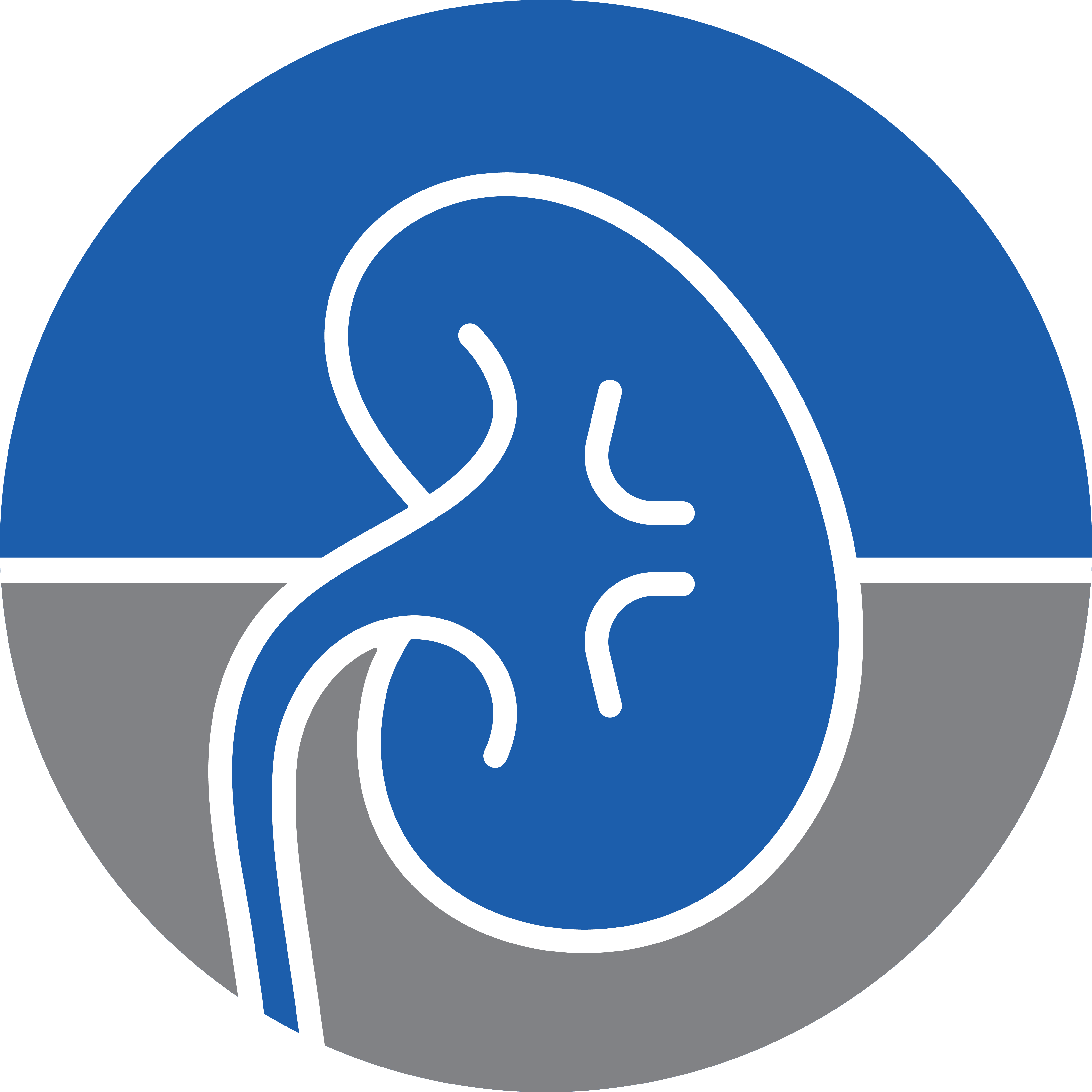
- Nephrology
- Neurology
- O&G and Peri-Natal
- Oncology
- Opthalmology
- Orthopedic
- Patient Support System
- Physio & Rehab
- Primary Care
- Respiratory Care
- Surgical Workplace
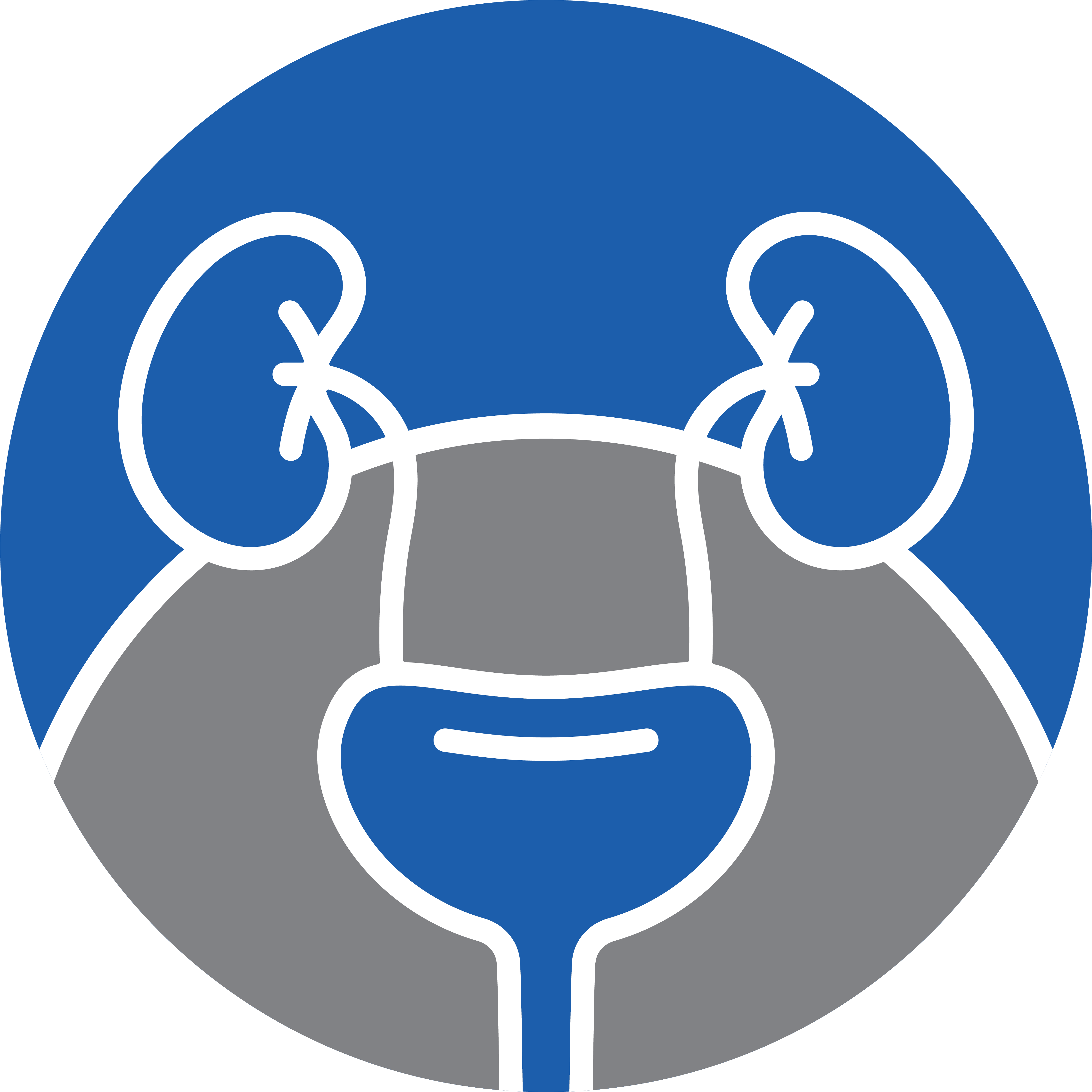
- Urology
- Wound Management
- Our Services
- Partners
- idsMED Learning Academy
- News
- Talent & Jobs
- Login / Sign Up